
Persistent hip pain can be more than just an inconvenience—it may be a sign of structural issues within the joint. Two common causes of hip discomfort, especially in active individuals and athletes, are cam and pincer lesions. The root of that discomfort may be a condition known as femoroacetabular impingement (FAI), which involves abnormal contact between the bones of the hip joint. Two key types of FAI—cam and pincer lesions—can limit mobility and, if left untreated, cause long-term joint damage. In this comprehensive guide, we’ll break down what cam and pincer lesions are, explore their symptoms, explain how they’re diagnosed, risk factors, and outline available treatment options, helping you take the necessary steps toward managing your hip health and returning to an active, pain-free lifestyle.
What Are Cam and Pincer Lesions?
Cam and pincer lesions are two distinct but related forms of femoroacetabular impingement (FAI), a condition where abnormal bone structures in the hip joint lead to increased friction and damage. Cam lesions occur when the femoral head (the ball of the hip joint) is not perfectly round, which interferes with smooth joint movement and causes the head to jam against the socket, damaging the cartilage and labrum. In contrast, pincer lesions arise when the hip socket (acetabulum) extends too far over the femoral head, leading to the labrum being compressed or “pinched,” particularly during hip flexion.
While the source of impingement differs—cam affecting the femur and pincer involving the acetabulum—the outcome is often similar: pain, reduced mobility, and joint deterioration. These lesions are frequently paired together because they often coexist, forming what’s known as combined FAI. When both types are present, the risk of joint damage and early hip osteoarthritis increases, underscoring the importance of timely diagnosis and treatment.
Symptoms of Hip Impingement
Cam and pincer lesions often present with noticeable discomfort and movement limitations, especially during activities that engage the hip joint. Common symptoms include:
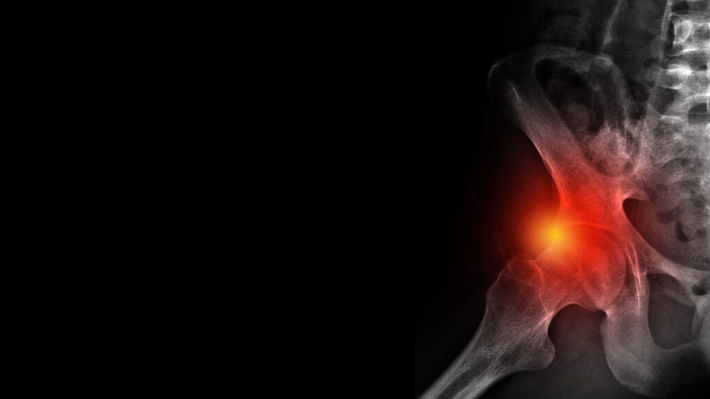
- Groin or Hip Pain: A persistent ache or sharp, stabbing pain felt deep in the front of the hip or groin area. This discomfort often worsens with physical activity, such as running or jumping, and may also intensify during or after prolonged sitting.
- Reduced Range of Motion: Stiffness or a noticeable decrease in the hip’s ability to move freely, making everyday actions like squatting, bending, or turning difficult. This limitation may be caused by the abnormal shape of the bones restricting joint movement.
- Clicking or Locking Sensation: A sensation of the hip catching, locking, or producing a clicking noise during certain movements. This can be a sign that the labrum, the cartilage lining the socket, is being pinched or damaged.
- Pain During Flexion or Rotation: Increased discomfort when the hip is flexed-such as when sitting, bringing the knee toward the chest, or rotating the leg inward or outward. These motions can trigger impingement between the bones, especially in sports or activities requiring frequent hip movement.
- Discomfort After Activity: A feeling of soreness, tightness, or stiffness in the hip that becomes more noticeable after physical activity, prolonged walking, or standing. This post-activity discomfort can be a signal of ongoing irritation within the joint.

Diagnosing Cam and Pincer Lesions
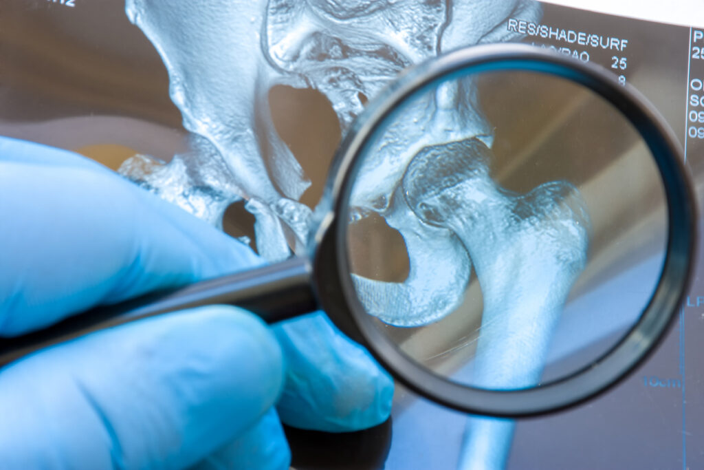
Accurately diagnosing cam and pincer lesions begins with a detailed evaluation of your symptoms and a physical exam to assess hip mobility, pain triggers, and range of motion. During the exam, your provider may perform specific movements to reproduce impingement pain and identify limitations. Imaging tests are essential for confirming the diagnosis. X-rays can reveal abnormal bone shapes or overgrowths on the femoral head (cam lesions) or acetabular rim (pincer lesions), while magnetic resonance imaging (MRI) may be used to assess soft tissue damage, such as labral tears or cartilage wear. In some cases, a computed tomography (CT) scan is recommended to provide more detailed images of the hip joint and guide treatment planning.
Risk Factors
Several factors can increase the likelihood of developing cam and pincer lesions. Genetics may play a role, as some individuals are born with structural differences in the hip joint that predispose them to femoroacetabular impingement. Abnormal bone development during adolescence—particularly during periods of rapid growth—can also lead to irregularities in the shape of the femoral head or acetabulum. High-impact sports that involve repetitive hip movement, such as soccer, hockey, or dance, place significant stress on the joint and are linked to a higher risk of developing FAI. Additionally, previous hip injuries or trauma can alter joint alignment and contribute to impingement over time. Regardless of the cause, early identification of these risk factors can help prevent long-term joint damage.
Treatment Options
There are several treatment strategies for managing cam and pincer lesions, depending on the severity of symptoms and how much joint damage is present. Below are five common approaches:
- Activity Modification: Adjusting your routine by avoiding movements that stress the hip joint—such as deep bending, squatting, or participating in high-impact sports like running or basketball—can help minimize strain and inflammation. Taking breaks from activities that exacerbate pain can give the joint time to heal and prevent further damage.
- Physical Therapy: A personalized rehabilitation plan guided by a physical therapist is essential in strengthening the muscles surrounding the hip joint. By improving joint stability, enhancing flexibility, and restoring range of motion, physical therapy can alleviate pain and help improve mobility, providing long-term benefits for hip health.
- Medications and Injections: Over-the-counter nonsteroidal anti-inflammatory drugs (NSAIDs), such as ibuprofen, can offer relief from pain and inflammation. In more severe cases, corticosteroid injections directly into the hip joint may be used to reduce swelling and provide longer-lasting pain relief, allowing individuals to better engage in physical therapy and other rehabilitation efforts.
- Hip Arthroscopy: For patients who don’t respond to conservative treatments, hip arthroscopy may be necessary. This minimally invasive surgical procedure involves using small incisions and a camera to visualize the joint. The surgeon can remove or reshape damaged bone, repair torn cartilage or the labrum, and improve joint function. Recovery time is typically shorter than with traditional surgery, making it a preferred option for many.
- Lifestyle Adjustments: Maintaining a healthy body weight is essential for reducing pressure on the hip joint, while incorporating low-impact exercises—such as swimming, cycling, or using an elliptical machine—can protect the joint from further irritation. Additionally, wearing supportive shoes and avoiding prolonged periods of standing can contribute to better hip health and overall comfort.
Contact Us
Cam and pincer lesions are common causes of hip pain that, if left unaddressed, can lead to long-term joint damage and reduced mobility. Fortunately, with proper diagnosis and treatment, most individuals can experience significant relief and return to the activities they enjoy. Whether you’re dealing with persistent discomfort or suspect an underlying issue like femoroacetabular impingement, seeking expert care is essential. At Peter Howard, M.D., we provide specialized evaluation and treatment for hip conditions, including arthroscopic repair for cam and pincer lesions. Schedule an appointment today to take the first step toward restoring your comfort and protecting your hip health.


